
A magazine where the digital world meets the real world.
On the web
- Home
- Browse by date
- Browse by topic
- Enter the maze
- Follow our blog
- Follow us on Twitter
- Resources for teachers
- Subscribe
In print
What is cs4fn?
- About us
- Contact us
- Partners
- Privacy and cookies
- Copyright and contributions
- Links to other fun sites
- Complete our questionnaire, give us feedback
Search:
Looking inside medicine - computer scientists in the body
Computer scientists are helping doctors, surgeons, biologists and psychologists get inside the body and mind, and improving the way that medical care will be provided now and in the future. It's a fascinating story of biology, maths and computing and it all starts with an X.
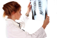
What a picture!
X-rays were the first practical method of examining the inside of a living body. The process involves firing high energy X-rays through the body with a photographic plate at the other side. Dense bits of the body like bones absorb radiation. That leads to a lighter area on the developed photographic negative. In effect a shadow is cast through you onto the photograph, giving a view inside. A problem with this is that, as with any camera, it's hard to get the photograph exposure right. Worse you have to find the space to store hundreds and thousands of sheets of film. Worse still, suppose your doctor in Manchester needs the X-ray taken of you when you are wanting to play football so you are in Frankfurt. The film has to be sent by post. Enter computer scientists to make things easier.
Portable pixel pictures
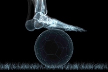
New digital X-ray systems are being developed. These use X-ray detectors not film and produce digital images rather than the standard photographic images. The advantage here is that those images can be processed using clever algorithms to correct for problems in exposure, or even to pick up particular shapes in the image. The diagnosis can be helped by the artificial intelligence in the computer, which can spot unusual patterns in the image and alert the doctor. Better still since these digital X-rays are computer based. They can be easily stored and transmitted throughout the world to places where they are needed.
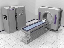
A slice of life
X-rays, even digital X-rays, can only give you flat images of the body innards. Like a shadow they squash all the depth details. Your insides are three-dimensional (3D) though, so it would be useful to be able to slice through your body and get a view inside. This is possible using a computer based method called tomography, from the Greek tomos (slice) and graphia (describing). It still uses X-rays but in tomography the X-ray source and the detector rotate round the body taking lots of images at different angles. It's like casting different shadows as the sun moves round you. So imagine you're using tomography on a cylinder, and your X-ray source is a torch. Move the torch round the cylinder and look at the shadow cast on a piece of paper moving at the opposite side to the torch. Each 'shadow' picture would look the same because a cylinder is circularly symmetric. Now imagine a more interesting shape. Each of the shadow pictures would depend on where you were at the time in relation to the shape. With some clever maths, a reconstruction algorithm and a computer you can go from the shadow pictures back to the shape. These shapes are the organs and innards of your body, and they can be recorded in their full 3D glory. There are now systems that spiral the X-ray source round the body making it quicker. You can even do tomography at very high speed allowing slices through the beating heart to be calculated. Interestingly the maths behind this technology, called the "Radon transform" after Czech mathematician Johann Radon (1887-1956), was developed purely as an abstract mathematical theory. No one at the time could see any use for it!
Check in at the Digital Hospital
Life saving healthcare and medical imaging is going digital. Using video conferencing, mobile scanners and even remote operated robotic surgery the field of tele-medicine allows expert medical care to be provided any time, any place. Today's progress towards the digital hospital combines different ways of taking information about the state of your body, such as digital X-rays, or tomographic images, readings from digital thermometers or digital blood pressure readers. We can combine all this information with your personal information into one big file, so there is no need for multiple paper copies to get out of date or lost. The hospital information system keeps track of all your data, and also importantly who has access to it.
Tomorrow's world and you
According to Alan McBride, a computer scientist who is working on these state of the art medical systems:

'This technology is a major step forward in health care where the UK is leading the way. The government's grand scheme will allow images taken in Newcastle to be shown on your GP's desk in London, together with the hospital report, which will automatically be emailed to their inbox. Computer science is playing the major role in all this, creating new ways to aid clinical practice, with plenty of scope in the future for talented computer scientists to get involved.'
The computer scientists who make this happen will not only be technical specialists but also experts in understanding human behaviour. We will only get the benefits such a grand scheme promises if the conflicting needs and concerns of all those involved are taken into account: patients, nurses, doctors, managers and politicians...that will take major people skills.